
Health Library
Retinal artery occlusion
Central retinal artery occlusion; CRAO; Branch retinal artery occlusion; BRAO; Vision loss - retinal artery occlusion; Blurry vision - retinal artery occlusion
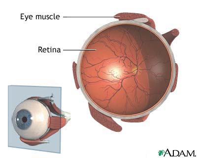
Retinal artery occlusion is a blockage in one of the small arteries that carry blood to the retina. The retina is a layer of tissue in the back of the eye that is able to sense light.
Images
I Would Like to Learn About:
Causes
Retinal arteries may become blocked when a blood clot or fat deposits get stuck in the arteries. These blockages are more likely if there is hardening of the arteries (atherosclerosis) in the eye.
Clots may travel from other parts of the body and block an artery in the retina. The most common sources of clots are the heart and carotid artery in the neck.
Most blockages occur in people with conditions such as:
- Carotid artery disease, in which the two large blood vessels in the neck become narrowed or blocked
- Diabetes
- Heart rhythm problem (mainly atrial fibrillation)
- Heart valve problem
- High levels of fat in the blood (hyperlipidemia)
- High blood pressure
- Intravenous drug use
- Temporal arteritis (inflammation of arteries due to an immune response)
If a branch of the retinal artery is blocked, part of the retina will not receive enough blood and oxygen. If this happens, you may lose part of your vision.
Symptoms
Sudden blurring or loss of vision may occur in:
- All of one eye (central retinal artery occlusion or CRAO)
- Part of one eye (branch retinal artery occlusion or BRAO)
The retinal artery occlusion may last for only a few seconds or minutes, or it may be permanent.
A blood clot in the eye may be a warning sign of clots elsewhere. A clot in the brain may cause a stroke.
Exams and Tests
Tests to evaluate the retina may include:
- Examination of the retina after dilating the pupil
- Fluorescein angiography
- Intraocular pressure
- Pupil reflex response
- Refraction
- Retinal photography
- Slit lamp examination
- Testing of side vision (visual field examination)
- Visual acuity
General tests should include:
- Blood pressure
- Blood tests, including cholesterol and triglyceride levels and the erythrocyte sedimentation rate
- Physical examination
Tests to identify the source of a clot from another part of the body may include:
- Echocardiogram
- Electrocardiogram (ECG)
- Heart monitor for abnormal heart rhythm
- Duplex Doppler ultrasound of the carotid arteries
Treatment
There is no proven treatment for vision loss that involves the whole eye, unless it is caused by another illness that can be treated.
Several treatments may be tried. To be helpful, these treatments must be given within 2 to 4 hours after symptoms begin. However, the benefit of these treatments has never been proven, and they are rarely used.
- Breathing in (inhaling) a carbon dioxide-oxygen mixture. This treatment causes the arteries of the retina to widen (dilate).
- Breathing in (inhaling) a high concentration of oxygen. This can help the retina survive, especially if the blockage is not complete.
- Massage of the eye.
- Removal of fluid from within the eye. An eye doctor uses a needle to drain a small amount of fluid from the front of the eye. This causes a sudden drop in eye pressure, which can sometimes cause the clot to move into a smaller branch artery where it will cause less damage.
- The clot-busting medicine, tissue plasminogen activator (tPA).
- Aspirin and other blood thinners.
The health care provider should look for the cause of the blockage. Blockages may be signs of a life-threatening medical problem.
Outlook (Prognosis)
People with blockages of the retinal artery may not get their vision back.
Possible Complications
Complications may include:
- Glaucoma (CRAO only)
- Partial or complete loss of vision in the affected eye
- Stroke (due to the same factors that contribute to retinal artery occlusion, not due to the occlusion itself)
When to Contact a Medical Professional
Contact your provider if you have sudden blurring or vision loss.
Prevention
Measures used to prevent other blood vessel (vascular) diseases, such as coronary artery disease, may decrease the risk for retinal artery occlusion. These include:
- Eating a low-fat diet
- Exercising
- Stopping smoking
- Losing weight if you are overweight
Sometimes, blood thinners may be used to prevent the artery from becoming blocked again. Aspirin or other anti-clotting medicines are used if the problem is in the carotid arteries. Warfarin or other more potent blood thinners are used if the problem is in the heart.
Related Information
RetinaBlood clots
Glaucoma
High blood pressure in adults - hypertension
Diabetes
Acquired platelet function defect
Atherosclerosis
High blood cholesterol levels
Vision problems
Blindness and vision loss
Stroke
References
Cioffi GA, Liebmann JM. Diseases of the visual system. In: Goldman L, Cooney KA, eds. Goldman-Cecil Medicine. 27th ed. Philadelphia, PA: Elsevier; 2024:chap 391.
Duker JS, Duker JS. Retinal arterial obstruction. In: Yanoff M, Duker JS, eds. Ophthalmology. 6th ed. Philadelphia, PA: Elsevier; 2023:chap 6.15.
Guluma K, Lee JE. Ophthalmology. In: Walls RM, ed. Rosen's Emergency Medicine: Concepts and Clinical Practice. 10th ed. Philadelphia, PA: Elsevier; 2023:chap 57.
Patel PS, Sadda SR, Sarraf D. Retinal artery and capillary occlusions. In: Sadda SR, Sarraf D, Freund KB, et al, eds. Ryan's Retina. 7th ed. Philadelphia, PA: Elsevier; 2023:chap 53.
Preterre C, Gaultier A, Obadia M, et al. Intravenous alteplase versus oral aspirin for acute central retinal artery occlusion within 4·5 h of severe vision loss (THEIA): a multicentre, double-dummy, patient-blinded and assessor-blinded, randomised, controlled, phase 3 trial. Lancet Neurol. 2025;24(11):909-919. PMID: 41109232 pubmed.ncbi.nlm.nih.gov/41109232/.
BACK TO TOPReview Date: 1/27/2026
Reviewed By: Franklin W. Lusby, MD, Ophthalmologist, Lusby Vision Institute, La Jolla, CA. Also reviewed by David C. Dugdale, MD, Medical Director, Brenda Conaway, Editorial Director, and the A.D.A.M. Editorial team.
 | A.D.A.M., Inc. is accredited by URAC, for Health Content Provider (www.urac.org). URAC's accreditation program is an independent audit to verify that A.D.A.M. follows rigorous standards of quality and accountability. A.D.A.M. is among the first to achieve this important distinction for online health information and services. Learn more about A.D.A.M.'s editorial policy, editorial process and privacy policy. A.D.A.M. is also a founding member of Hi-Ethics. This site complies with the HONcode standard for trustworthy health information: verify here. |
The information provided herein should not be used during any medical emergency or for the diagnosis or treatment of any medical condition. A licensed medical professional should be consulted for diagnosis and treatment of any and all medical conditions. Links to other sites are provided for information only -- they do not constitute endorsements of those other sites. No warranty of any kind, either expressed or implied, is made as to the accuracy, reliability, timeliness, or correctness of any translations made by a third-party service of the information provided herein into any other language.
© 1997-
2026 A.D.A.M., a business unit of Ebix, Inc. Any duplication or distribution of the information contained herein is strictly prohibited.
All content on this site including text, images, graphics, audio, video, data, metadata, and compilations is protected by copyright and other intellectual property laws. You may view the content for personal, noncommercial use. Any other use requires prior written consent from Ebix. You may not copy, reproduce, distribute, transmit, display, publish, reverse-engineer, adapt, modify, store beyond ordinary browser caching, index, mine, scrape, or create derivative works from this content. You may not use automated tools to access or extract content, including to create embeddings, vectors, datasets, or indexes for retrieval systems. Use of any content for training, fine-tuning, calibrating, testing, evaluating, or improving AI systems of any kind is prohibited without express written consent. This includes large language models, machine learning models, neural networks, generative systems, retrieval-augmented systems, and any software that ingests content to produce outputs. Any unauthorized use of the content including AI-related use is a violation of our rights and may result in legal action, damages, and statutory penalties to the fullest extent permitted by law. Ebix reserves the right to enforce its rights through legal, technological, and contractual measures.
